

The field of view is tailored to you
We scan the area the clinical question needs, and no more. The choice is made with the referring clinician, following the principles of justification and optimisation.
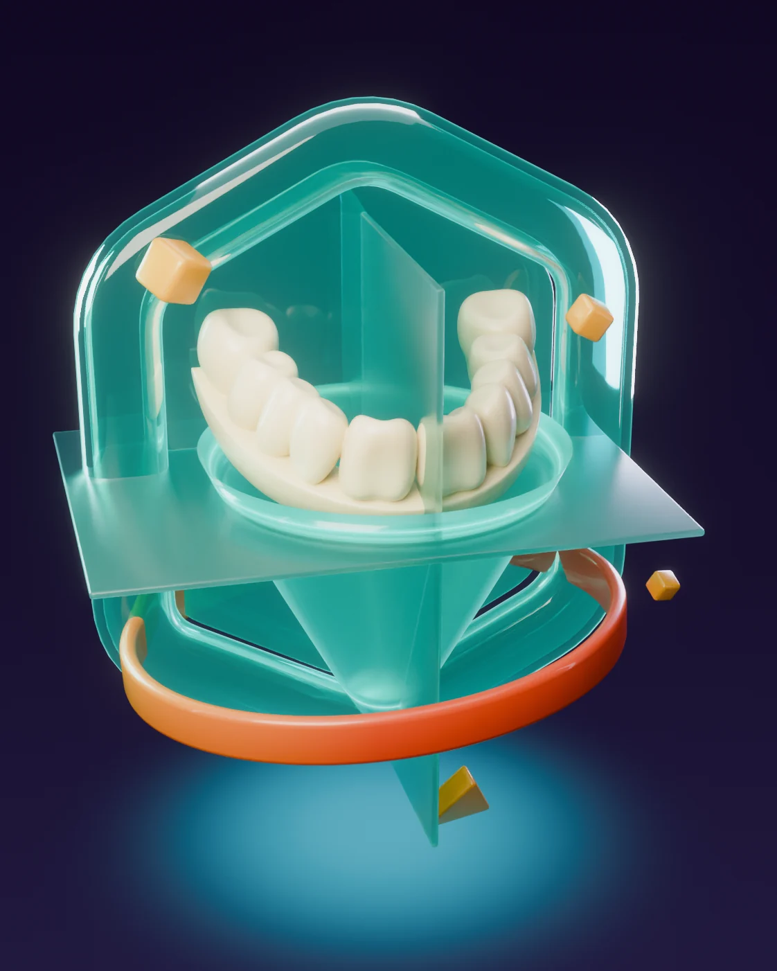
A three-dimensional image of the dental and maxillofacial structures, for when a two-dimensional view cannot answer the clinical question well enough.
A CBCT builds a three-dimensional dataset from many X-ray projections, so the area can be looked at from every side. The field of view and the protocol are chosen for the question on the referral. It is not a routine test, and it is not automatically “better” than the right two-dimensional image.
We scan the area the clinical question needs, and no more. The choice is made with the referring clinician, following the principles of justification and optimisation.
The machine turns around your head and collects many projections. Software reconstructs them into cross-sections and three-dimensional data that can be reviewed in different planes.
Depending on the referral, this may include images, DICOM data and a written radiology report. The report supports the professional looking after you. It is not a treatment plan.
Movement, metal restorations and other materials can create artefacts in the image. CBCT mainly shows hard tissue, bone and teeth, so it is not the right method for every soft-tissue question.
The images and, where a report has been requested, the report reach you and your referring clinician securely.
A report is written where one has been requested, and it supports the professional looking after you. It is not a treatment plan.Selected guidelines and publications related to the content of this page. Each link opens the publication on the relevant organisation’s website.
Request the examination on your referral or contact our team about working with DentalRay and referring patients.