


For patients
Bring the referral or the instructions from your dentist. If you are not sure which examination was requested, call us before you come and we will work it out together.
Eight examinations, from the simple panoramic to the 3D scan. The one you will have is written on your referral: see here what to expect from each.
Every shape is an examination. Choose one to learn more.
Panoramic X-ray
One image that shows your teeth, your jaws and the neighbouring structures together. It is used when a broad overview is what is needed.
Cone-Beam Computed Tomography (CBCT)
A three-dimensional image of the dental and maxillofacial structures, for when a two-dimensional view cannot answer the clinical question well enough.
Full Mouth - complete series of intraoral X-rays
A set of small, targeted intraoral views that shows each region in more detail than a panoramic overview can.
Periapical Intraoral X-Ray
A detailed intraoral view of one or a few teeth, from the crown down to the tip of the root and the bone around it.
Bitewing X-Ray
An intraoral view that shows the crowns of the upper and lower back teeth together, along with the alveolar bone in the same region.
Cephalometric X-Ray
A standardised side or front-on image of the head, used mainly for orthodontic analysis and for following change over time.
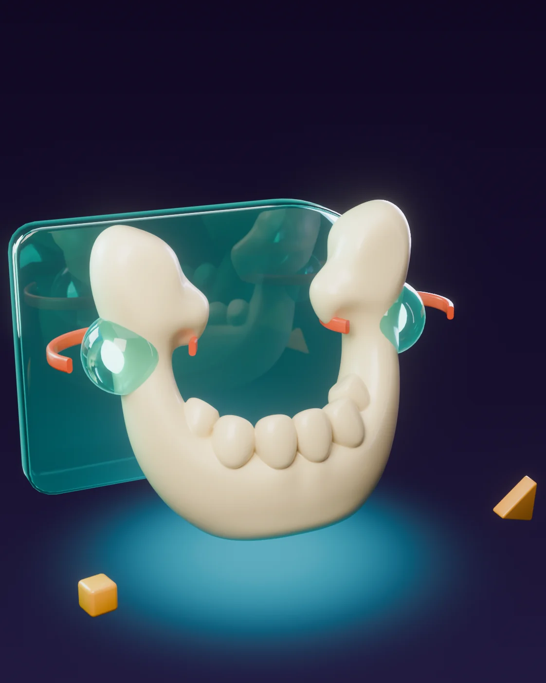
Temporomandibular Joints (TMJ)
Targeted imaging of the bone in the temporomandibular joints, where the lower jaw meets the skull. The protocol follows the clinical question.
Occlusal X-Ray
An intraoral view taken with a larger plate, covering a wider area of the upper or lower jaw for specific clinical questions.
Your dentist chooses the examination and writes it on your referral.
Some examinations show the whole mouth at a glance, others focus on a single tooth or see in three dimensions. We follow what your referral says and confirm the area with you before the examination.
Bring the referral or the instructions from your dentist. If you are not sure which examination was requested, call us before you come and we will work it out together.
A clear clinical question, the region of interest and any relevant previous imaging let us select and optimise the acquisition protocol.
Dental imaging helps the attending dentist to distinguish diseases in the teeth and the surrounding structures and tissues, which cannot be distinguished only clinically. In addition, it helps the dentist to find and treat dental problems at an early stage, thus protecting the patient from additional costs and possible progression of the disease. These images are divided into the following categories:
Request the examination on your referral or contact our team about working with DentalRay and referring patients.