

Lateral and posteroanterior views
The lateral view shows, from the side, how the skull, the jaws and the teeth relate to each other. A posteroanterior view is requested when a front-on image and a side-to-side comparison are needed.
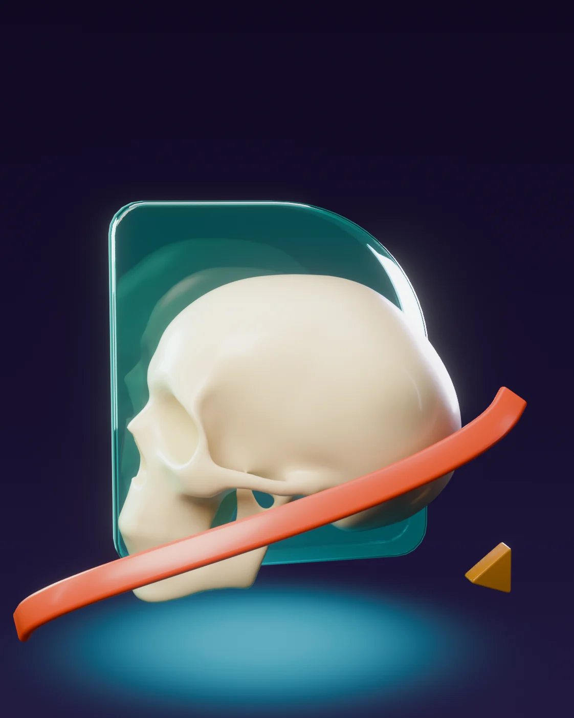
A standardised side or front-on image of the head, used mainly for orthodontic analysis and for following change over time.
A cephalometric image records your skull, jaws and teeth with fixed, standardised geometry, so your orthodontist can take measurements and compare them over time. The image provides the data. The orthodontic diagnosis and the plan stay with your treating clinician.
The lateral view shows, from the side, how the skull, the jaws and the teeth relate to each other. A posteroanterior view is requested when a front-on image and a side-to-side comparison are needed.
The fixed, standardised position is what makes the measurements reliable and comparable. Our team steadies your head carefully and gives you specific instructions for your bite and your posture.
The standardised image reaches you and the orthodontist who referred you, digitally.
A report is written where one has been requested, and it supports the professional looking after you. It is not a treatment plan.Selected guidelines and publications related to the content of this page. Each link opens the publication on the relevant organisation’s website.
Request the examination on your referral or contact our team about working with DentalRay and referring patients.